Metaheuristic Analysis of Silicone Rubber Eyeshield on CT Image Quality in Head CT Examination

Downloads
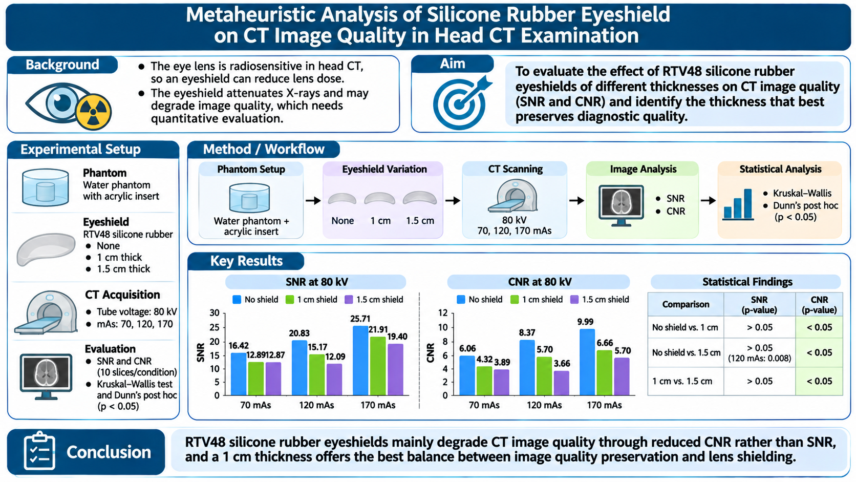
The eye lens is one of the most radiosensitive organs during head computed tomography (CT) examinations. Eyeshields can reduce lens dose, but they attenuate X-rays and may degrade image quality, so their effect on quantitative image quality parameters requires further investigation. This study evaluated the effect of RTV48 silicone rubber eyeshields of different thicknesses on CT image quality and identified the thickness that best preserves diagnostic quality. A water phantom with an acrylic insert was scanned without an eyeshield, with a 1 cm eyeshield, and with a 1.5 cm eyeshield. Acquisitions used a constant tube voltage of 80 kV and tube current-time products of 70, 120, and 170 mAs. Image quality was assessed quantitatively using signal-to-noise ratio (SNR) and contrast-to-noise ratio (CNR) from ten image slices per condition. Statistical analysis used the Kruskal–Wallis test followed by Dunn’s post hoc test at a significance level of p < 0.05. Increasing mAs generally improved SNR and CNR in all conditions, whereas the eyeshields reduced both parameters through photon attenuation, with larger reductions at greater thickness. Most SNR differences between the no-eyeshield and eyeshield conditions were not statistically significant (p > 0.05), except for the 1.5 cm eyeshield at 120 mAs (p = 0.008). In contrast, all CNR comparisons differed significantly (p < 0.05), indicating that eyeshields affect image contrast more strongly than noise characteristics. The 1 cm eyeshield preserved image quality better than the 1.5 cm eyeshield. These findings show that RTV48 silicone rubber eyeshields mainly degrade CT image quality through reduced CNR rather than SNR, and that a 1 cm thickness offers the best balance between image quality preservation and lens shielding
[1] E. C. Lin, “Radiation risk from medical imaging,” Mayo Clin. Proc., vol. 85, no. 12, pp. 1142–1146, 2010, doi: 10.4065/mcp.2010.0260.
[2] H. Alsleem and R. Davidson, “Factors affecting contrast-detail performance in computed tomography: A review,” J. Med. Imaging Radiat. Sci., vol. 44, no. 2, pp. 62–70, 2013, doi: 10.1016/j.jmir.2012.12.001.
[3] J. Tarkiainen, M. Nadhum, A. Heikkilä, I. Rinta-Kiikka, and A. Joutsen, “Radiation dose of the eye lens in CT examinations of the brain in clinical practice—the effect of radiographer training to optimise gantry tilt and scan length,” Radiat. Prot. Dosimetry, vol. 199, no. 5, pp. 391–398, 2023, doi: 10.1093/rpd/ncad002.
[4] S. Poosiri, K. Chuboonlap, and N. Kaewlaied, “An Age-Based Size-Specific Dose Estimate for Pediatric Computed Tomography Head Examinations Performed at Songklanagarind Hospital, Thailand, from 2017 to 2019,” Appl. Sci., vol. 14, no. 17, Art. no. 7848, 2024, doi: 10.3390/app14177848.
[5] N. Hamada, T. V. Azizova, and M. P. Little, “An update on effects of ionizing radiation exposure on the eye,” Br. J. Radiol., vol. 93, no. 1115, Art. no. 20190829, 2020, doi: 10.1259/bjr.20190829.
[6] E. Della Vecchia et al., “Risk of cataract in health care workers exposed to ionizing radiation: A systematic review,” Med. Lav., vol. 111, no. 4, pp. 269–284, 2020, doi: 10.23749/mdl.v111i4.9045.
[7] M. P. Little, A. Patel, N. Hamada, and P. Albert, “Analysis of Cataract in Relationship to Occupational Radiation Dose Accounting for Dosimetric Uncertainties in a Cohort of U.S. Radiologic Technologists,” Radiat. Res., vol. 194, no. 2, pp. 153–161, 2020, doi: 10.1667/RR15529.1.
[8] J. J. Lucido III et al., “Development of transparent eye shields for total skin electron beam radiotherapy,” J. Appl. Clin. Med. Phys., vol. 23, no. 9, Art. no. e13722, 2022, doi: 10.1002/acm2.13722.
[9] A. Manuscript et al., “Ac ce pte d M us pt,” Mater. Today Proc., vol. 27, no. xxxx, pp. 0–31, 2019, [Online]. Available: https://doi.org/10.1016/j.matpr.2019.12.188%0Ahttps://doi.org/10.1016/j.matpr.2019.09.090%0Ahttps://doi.org/10.1080/14484846.2018.1432089
[10] N. J. AbuAlRoos, N. A. Baharul Amin, and R. Zainon, “Conventional and new lead-free radiation shielding materials for radiation protection in nuclear medicine: A review,” Radiat. Phys. Chem., vol. 165, Art. no. 108439, 2019, doi: 10.1016/j.radphyschem.2019.108439.
[11] A. Trajkovska Petkoska, “Assessment of the Attenuation Properties of Commercial Lead-Free Radiation-Shielding Composite Materials Against Medical X-rays,” J. Compos. Sci., vol. 7, no. 10, Art. no. 424, 2023, doi: 10.3390/jcs7100424.
[12] A. M. El-Khatib, E. Elesh, M. S. Hamada, E. M. Sabry, and M. M. Gouda, “Study Attenuation Parameters and Physical Properties of Silicone Rubber Reinforced with Nano- and Micro-Sized Aluminum Oxide Composites,” Silicon, vol. 16, no. 6, pp. 2621–2634, 2024, doi: 10.1007/s12633-024-02847-7.
[13] A. M. El-Khatib, K. Zard, M. I. Abbas, and M. M. Gouda, “Novel composite based on silicone rubber and a nano mixture of SnO₂, Bi₂O₃, and CdO for gamma radiation protection,” Sci. Rep., vol. 14, no. 1, Art. no. 1578, 2024, doi: 10.1038/s41598-024-51965-0.
[14] A. Saeed and W. A. Abu-raia, “Silicone rubber composite reinforced by bismuth tungsten oxide as an effective gamma ray protective materials,” J. Polym. Res., vol. 29, Art. no. 208, 2022, doi: 10.1007/s10965-022-03055-w.
[15] T. Yoshida, K. Sasaki, Y. Kawasaki, T. Hayakawa, T. Kawadai, and T. Shibasaki, “Evaluation of the dose reduction effect of crystalline lens exposure in cone-beam computed tomography with bismuth eye shield for image-guided radiation therapy: An anthropomorphic phantom study,” J. Appl. Clin. Med. Phys., vol. 26, no. 5, Art. no. e70024, 2025, doi: 10.1002/acm2.70024.
[16] H. Kosaka et al., “Radiation dose reduction to the eye lens in head CT using tungsten functional paper and organ-based tube current modulation,” Eur. J. Radiol., vol. 124, no. November 2019, p. 108814, 2020, doi: 10.1016/j.ejrad.2020.108814.
[17] Y. L. Liao, N. K. Lai, Y. S. Tyan, and H. Y. Tsai, “Bismuth shield affecting CT image quality and radiation dose in adjacent and distant zones relative to shielding surface: A phantom study,” Biomed. J., vol. 42, no. 5, pp. 343–351, 2019, doi: 10.1016/j.bj.2019.04.004.
[18] Y. H. Lee, S. H. Yang, Y. K. Lin, R. D. Glickman, C. Y. Chen, and W. P. Chan, “Eye Shielding During Head CT Scans: Dose Reduction and Image Quality Evaluation,” Acad. Radiol., vol. 27, no. 11, pp. 1523–1530, 2020, doi: 10.1016/j.acra.2019.12.011.
[19] F. S. Santos, P. C. Santana, and A. P. Mourão, “Comparative Study of the Use of Bismuth Shielding on Head Computed Tomography Scans Using Phantoms,” Braz. J. Radiat. Sci., vol. 8, no. 1B, pp. 1–10, 2020, doi: 10.15392/bjrs.v8i1B.936.
[20] T. Pengpan, N. Rattanarungruangchai, J. Dechjaithat, P. Panthim, P. Siricharuwong, and A. Prapan, “Optimization of Image Quality and Organ Absorbed Dose for Pediatric Chest X-Ray Examination: In-House Developed Chest Phantom Study,” Radiol. Res. Pract., vol. 2022, pp. 1–10, 2022, doi: 10.1155/2022/3482458.
[21] G. Pahn, S. Skornitzke, H. P. Schlemmer, H. U. Kauczor, and W. Stiller, “Toward standardized quantitative image quality (IQ) assessment in computed tomography (CT): A comprehensive framework for automated and comparative IQ analysis based on ICRU Report 87,” Phys. Medica, vol. 32, no. 1, pp. 104–115, 2016, doi: 10.1016/j.ejmp.2015.09.017.
[22] A. E. Michael et al., “Nonenhanced Photon Counting CT of the Head: Impact of the keV Level, Iterative Reconstruction and Calvaria on Image Quality in Monoenergetic Images,” Clin. Neuroradiol., vol. 34, no. 1, pp. 75–83, 2024, doi: 10.1007/s00062-023-01331-w.
[23] N. E. Wulandari, H. Sutanto, C. Anam, H. Sofyan, and A. H. Purbarani, “A Comparative Analysis between SR-Pb Shield and TCM in Eye Dose of Head CT Examination,” Int. J. Sci. Res. Sci. Technol., vol. 10, no. 6, pp. 408–413, 2023, doi: 10.32628/IJSRST52310683.
[24] H. M. Y. Okda, E. R. Sheha, F. Zahran, M. A. Yousef, T. I. Shaheen, and A. G. Hassabo, “Development of multilayered polymer-BaSO4 composites for flexible and efficient lead-free X-ray shielding,” Sci. Rep., vol. 16, no. 1, p. 6719, 2026, doi: 10.1038/s41598-026-37398-x.
[25] E. Hidayanto et al., “Effect of variation of silicone rubber RTV 52 and bluesil catalyst 60 R composition on bolus material for electron beam radiotherapy application,” Biomed. Phys. Eng. Express, vol. 8, no. 4, Art. no. 045005, 2022, doi: 10.1088/2057-1976/ac6f24.
[26] M. Dieckmeyer et al., “Computed Tomography of the Head: A Systematic Review on Acquisition and Reconstruction Techniques to Reduce Radiation Dose,” Clin. Neuroradiol., vol. 33, no. 3, pp. 591–610, 2023, doi: 10.1007/s00062-023-01271-5.
[27] V. Karami et al., “Tradeoffs between Radiation Exposure to the Lens of the Eyes and Diagnostic Image Quality in Pediatric Brain Computed Tomography,” J. Med. Signals Sens., vol. 13, no. 3, pp. 208–216, 2023, doi: 10.4103/jmss.jmss_19_22.
[28] A. Safari, P. Rafie, S. Taeb, M. Najafi, and S. M. J. Mortazavi, “Development of Lead-Free Materials for Radiation Shielding in Medical Settings: A Review,” J. Biomed. Phys. Eng., vol. 14, no. 3, pp. 229–244, 2024, doi: 10.31661/jbpe.v0i0.2404-1742.
[29] A. M. El, K. E. Elesh, M. S. Hamada, E. M. Sabry, and M. M. Gouda, “Study Attenuation Parameters and Physical Properties of Silicone Rubber Reinforced with Nano ‑ and Micro ‑ Sized Aluminum Oxide Composites,” Silicon, pp. 2621–2634, 2024, doi: 10.1007/s12633-024-02847-7.
[30] K. Igarashi, K. Imai, S. Matsushima, C. Yamauchi-Kawaura, and K. Fujii, “Development and validation of the effective CNR analysis method for evaluating the contrast resolution of CT images,” Phys. Eng. Sci. Med., vol. 47, no. 2, pp. 717–727, 2024, doi: 10.1007/s13246-024-01400-5.
[31] M. Pula, E. Kucharczyk, A. Zdanowicz, and M. Guzinski, “Image Quality Improvement in Deep Learning Image Reconstruction of Head Computed Tomography Examination,” Tomography, vol. 9, no. 4, pp. 1485–1493, 2023, doi: 10.3390/tomography9040118.
[32] M. M. Woeltjen et al., “Low-Dose High-Resolution Photon-Counting CT of the Lung : Radiation Dose and Image Quality in the Clinical Routine,” 2022.
[33] F. S. Maddaloni et al., “Assessment of Image Quality Performance of a Photon-Counting Computed Tomography Scanner Approved for Whole-Body Clinical Applications,” Sensors, vol. 25, no. 23, Art. no. 7338, 2025, doi: 10.3390/s25237338.
[34] C. H. Kristiansen et al., “Halved contrast medium dose coronary dual-layer CT-angiography – phantom study of tube current and patient characteristics,” Int. J. Cardiovasc. Imaging, vol. 40, no. 4, pp. 931–940, 2024, doi: 10.1007/s10554-024-03062-6.
[35] F. Zahroh, C. Anam, H. Sutanto, Y. Irdawati, Z. Arifin, and Y. Kartikasari, “Effect of Silicone Rubber-Lead (SR-Pb) Thickness on Dose Reduction and Image Quality as Gonad Shield,” J. Biomed. Phys. Eng., vol. 10, no. 6, pp. 699–706, 2020, doi: 10.31661/jbpe.v0i0.1912-1007.
[36] A. Banaei, A. Dadashi, S. S. Zakariaee, and V. Saba, “Assessing the image quality and eye lens dose reduction using bismuth shielding in computed tomography of brain,” J. Kerman Univ. Med. Sci., vol. 25, no. 6, pp. 471–482, 2018.
[37] Y. Irdawati, H. Sutanto, C. Anam, E. Hidayanto, F. Zahroh, G. Wiratma Jaya, and B. Susanto, “Silicone rubber with lead-acid composite as alternative radiation filter in digital radiography (DR),” J. Phys.: Conf. Ser., vol. 1505, no. 1, Art. no. 012035, 2020, doi: 10.1088/1742-6596/1505/1/012035.
[38] H. R. S. Larjava, C. T. M. Eneh, and H. M. Niiniviita, “To shield or not to shield: shielding may have unintended effects on patient dose in CT,” Eur. Radiol., vol. 34, no. 4, pp. 2480–2486, 2024, doi: 10.1007/s00330-023-10211-3.
[39] Y. Liao, N. Lai, Y. Tyan, and H. Tsai, “ScienceDirect Bismuth shield affecting CT image quality and radiation dose in adjacent and distant zones relative to shielding surface : A phantom study,” Biomed. J., vol. 42, no. 5, pp. 343–351, doi: 10.1016/j.bj.2019.04.004.
[40] T. Pengpan, N. Rattanarungruangchai, J. Dechjaithat, P. Panthim, P. Siricharuwong, and A. Prapan, “Optimization of Image Quality and Organ Absorbed Dose for Pediatric Chest X-Ray Examination : In-House Developed Chest Phantom Study,” vol. 2022, 2022.
[41] [41] T. Prabsattroo, K. Wachirasirikul, P. Tansangworn, P. Punikhom, and W. Sudchai, “The Dose Optimization and Evaluation of Image Quality in the Adult Brain Protocols of Multi-Slice Computed Tomography: A Phantom Study,” J. Imaging, vol. 9, no. 12, Art. no. 264, 2023, doi: 10.3390/jimaging9120264.
[42] F. Elkut et al., “The Influence of mAs on Effective Dose and Image Quality in Chest CT scans Using RANDO Phantom,” Libyan J. Med. Appl. Sci., vol. 3, no. 2, pp. 53–58, 2025, doi: 10.64943/ljmas.v3i2.64.
[43] C. Anam, R. Amilia, A. Naufal, T. Fujibuchi, and G. Dougherty, “A statistical-based automatic detection of a low-contrast object in the ACR CT phantom for measuring contrast-to-noise ratio of CT images,” Biomed. Phys. Eng. Express, vol. 11, no. 1, pp. 1–12, 2025, doi: 10.1088/2057-1976/ad90e9.
Copyright (c) 2026 NASYWA ALIFIA HANUM, Irmayatul Hikmah, Dodi Zulherman (Author)

This work is licensed under a Creative Commons Attribution-ShareAlike 4.0 International License.
Authors who publish with this journal agree to the following terms:
- Authors retain copyright and grant the journal right of first publication with the work simultaneously licensed under a Creative Commons Attribution-ShareAlikel 4.0 International (CC BY-SA 4.0) that allows others to share the work with an acknowledgement of the work's authorship and initial publication in this journal.
- Authors are able to enter into separate, additional contractual arrangements for the non-exclusive distribution of the journal's published version of the work (e.g., post it to an institutional repository or publish it in a book), with an acknowledgement of its initial publication in this journal.
- Authors are permitted and encouraged to post their work online (e.g., in institutional repositories or on their website) prior to and during the submission process, as it can lead to productive exchanges, as well as earlier and greater citation of published work (See The Effect of Open Access).